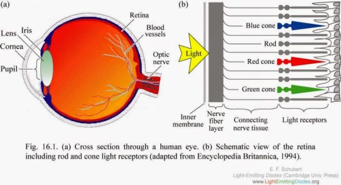
Embark on a journey into the realm of cones counterparts in the eye, where we uncover the fascinating structures that work hand in hand with cones to orchestrate our visual perception. Prepare to be captivated as we delve into their intricate functions and the profound impact they have on our ability to see and interpret the world around us.
From the distribution of cones in the retina to their remarkable adaptation to light and dark, we’ll explore the mechanisms that enable us to perceive color, distinguish fine details, and navigate our surroundings with ease.
Types of Cones
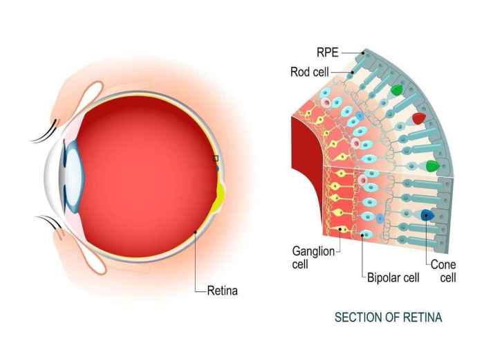
The human eye contains three types of cones, each sensitive to a specific range of wavelengths of light. These cones are responsible for our ability to perceive color.
The three types of cones are:
- Short-wavelength (S) cones:These cones are sensitive to wavelengths of light between 400 and 450 nanometers (nm), which corresponds to the color blue.
- Medium-wavelength (M) cones:These cones are sensitive to wavelengths of light between 500 and 570 nm, which corresponds to the color green.
- Long-wavelength (L) cones:These cones are sensitive to wavelengths of light between 570 and 650 nm, which corresponds to the color red.
The different types of cones work together to create our perception of color. When light enters the eye, it is focused on the retina, where the cones are located. The cones then send signals to the brain, which interprets the signals to create an image of the world around us.
For example, when we look at a green apple, the M cones in our eyes are most strongly stimulated. The brain interprets this signal as green, and we perceive the apple as green.
Cone Distribution in the Retina: Cones Counterparts In The Eye
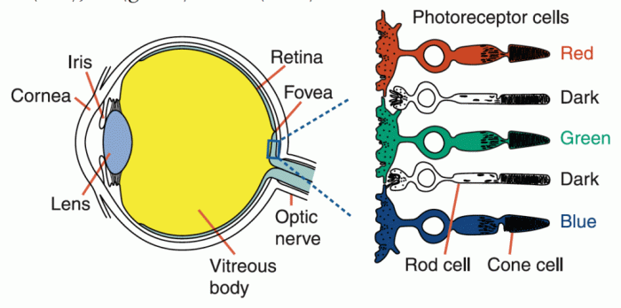
The distribution of cones in the retina is not uniform. There are far more cones in the central region of the retina, called the fovea, than in the peripheral regions. This distribution is responsible for our ability to see fine details in the center of our vision and to perceive colors more accurately in the central region.
In the realm of vision, the cones, our eyes’ counterparts to rods, serve as the maestros of color perception. While we delve into the intricate workings of the eye, let’s take a momentary detour to explore the intricacies of algebra 2 saxon 3rd edition . Returning to our ocular symphony, the cones, with their specialized pigments, allow us to perceive the vibrant tapestry of the world around us, enriching our visual experience with a kaleidoscope of hues.
Fovea
The fovea is a small, highly specialized area of the retina that contains only cones. It is located in the center of the macula, which is the area of the retina responsible for central vision. The fovea is responsible for our sharpest vision and our ability to perceive fine details.
It contains a high density of cones, which allows us to see objects in great detail. The fovea is also responsible for our ability to perceive colors accurately. It contains a high proportion of blue-sensitive cones, which are responsible for our ability to see blue and violet colors.
Peripheral Vision
The peripheral regions of the retina contain a lower density of cones than the fovea. This is because the peripheral regions are responsible for our peripheral vision, which is less detailed and less sensitive to color than central vision. The peripheral regions contain a higher proportion of rod cells, which are more sensitive to low levels of light than cones.
This allows us to see in low-light conditions, but with less detail and less color perception.
Visual Acuity and Color Perception
The distribution of cones in the retina plays a major role in our visual acuity and color perception. The high density of cones in the fovea gives us sharp vision and allows us to see fine details. The lower density of cones in the peripheral regions gives us less detailed vision and less color perception.
This distribution of cones allows us to see objects in great detail in the center of our vision, while still being able to see objects in our peripheral vision, even in low-light conditions.
Visual Field Size
The distribution of cones in the retina also affects our visual field size. The visual field is the area of space that we can see when we look straight ahead. The fovea is responsible for our central vision, which is the area of space that we can see most clearly.
The peripheral regions of the retina are responsible for our peripheral vision, which is the area of space that we can see around the central vision. The distribution of cones in the retina allows us to have a wide visual field, which allows us to see objects in front of us, as well as objects to the side of us.
Cone Function and Adaptation
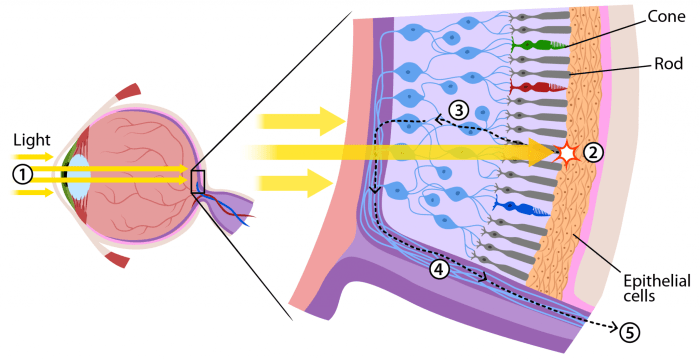
Cones are responsible for color vision and high-acuity vision in bright light conditions. They contain photopigments that absorb light and trigger electrical signals in the retina, which are then transmitted to the brain for visual processing.
Photopigments and Color Vision
Cones contain three types of photopigments, each sensitive to a specific wavelength of light:
- S-cones (short-wavelength): Sensitive to blue light (420-440 nm).
- M-cones (medium-wavelength): Sensitive to green light (530-550 nm).
- L-cones (long-wavelength): Sensitive to red light (560-580 nm).
The combination of signals from these three cone types allows us to perceive a wide range of colors.
Cone Adaptation
Cones adapt to changes in light intensity through two main mechanisms:
- Light Adaptation: When exposed to bright light, cones become less sensitive to reduce the risk of overstimulation. This process involves decreasing the amount of photopigment available for light absorption.
- Dark Adaptation: When exposed to dim light, cones become more sensitive to enhance vision in low-light conditions. This process involves increasing the amount of photopigment available for light absorption.
Dark adaptation can take up to 30 minutes to complete, which is why it takes time for our vision to adjust when moving from a bright to a dark environment.
Examples of Cone Adaptation
- When entering a dark room from a brightly lit area, it takes time for our eyes to adjust and see clearly due to the need for dark adaptation.
- When transitioning from dim light to bright sunlight, our eyes initially experience glare and reduced visual acuity as the cones adapt to the increased light intensity.
Cone Counterparts in the Eye
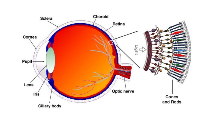
In the realm of vision, cones are the photoreceptor cells responsible for detecting color and fine details in bright light conditions. However, they do not work in isolation; they have counterparts that play equally important roles in the visual process.
Rods
Rods are the counterparts to cones and are specialized for low-light conditions. They are highly sensitive to light, allowing us to perceive shapes and movement even in dim environments. Unlike cones, rods are not sensitive to color and provide only grayscale vision.
The distribution of rods in the retina differs from that of cones. Rods are more concentrated in the peripheral regions, enabling us to have better night vision in the outer areas of our field of view.
In terms of function, rods and cones work in conjunction to provide a comprehensive visual experience. Cones are responsible for color vision and fine details in bright light, while rods take over in low-light conditions, allowing us to navigate and perceive objects in darkness.
Clinical Significance of Cones
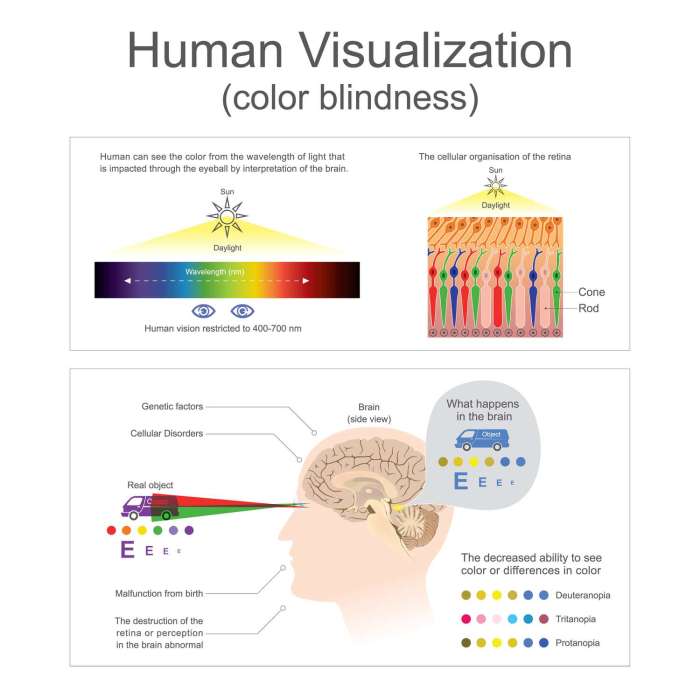
Cones, specialized photoreceptor cells in the eye, play a crucial role in our ability to perceive color and fine details. However, various disorders and diseases can affect cones, leading to impaired vision and color perception.
Common Disorders and Diseases Affecting Cones
Cone-related disorders and diseases can arise from genetic mutations, aging, or other factors. Some common examples include:
- Color Vision Deficiencies:These conditions, such as protanopia (red-blindness) and deuteranopia (green-blindness), result from defective or missing cone pigments.
- Cone Dystrophies:Inherited or acquired disorders that gradually damage cone photoreceptors, leading to progressive vision loss and color perception impairments.
- Macular Degeneration:Age-related degeneration of the central retina, which contains the highest concentration of cones, causing blurred vision and difficulty with color discrimination.
- Retinitis Pigmentosa:A group of inherited retinal diseases that affect both rods and cones, leading to night blindness, peripheral vision loss, and color vision impairments.
Impact of Cone Dysfunction on Vision and Color Perception
Dysfunction or loss of cones can significantly impact vision and color perception:
- Reduced Visual Acuity:Cones are responsible for sharp central vision, so cone dysfunction can lead to blurred vision, making it difficult to recognize details.
- Color Vision Deficiencies:Defective or missing cone pigments impair the ability to distinguish certain colors, leading to color blindness or reduced color perception.
- Night Vision Problems:Cones are less sensitive to low light conditions, so cone dysfunction can exacerbate night blindness.
Treatment Options and Management Strategies, Cones counterparts in the eye
Treatment options for cone-related disorders vary depending on the specific condition and its severity. Some approaches include:
- Corrective Lenses:Color-tinted lenses or filters can enhance color perception in individuals with color vision deficiencies.
- Low Vision Aids:Magnifying devices and other assistive technologies can improve visual acuity for individuals with cone dystrophies.
- Genetic Counseling:For inherited cone disorders, genetic counseling can help families understand the risks and options for managing the condition.
- Research and Clinical Trials:Ongoing research and clinical trials aim to develop new therapies and treatments for cone-related disorders.
Top FAQs
What are the counterparts of cones in the eye?
The counterparts of cones in the eye are specialized structures known as rods, horizontal cells, and bipolar cells.
How do cones and their counterparts work together?
Cones are responsible for color vision and high-acuity vision, while rods are responsible for low-light vision. Horizontal cells and bipolar cells transmit signals from cones to other neurons in the retina, enabling the processing of visual information.
What is the clinical significance of understanding cones and their counterparts?
Understanding cones and their counterparts is essential for diagnosing and treating visual disorders such as color blindness, night blindness, and macular degeneration.
